Ex Vivo Connectomics
Ex Vivo Connectomics is a postmortem imaging and histopathological study of brain specimens from patients who die from severe traumatic brain injury. The primary goal of this study is to identify the circuits within injured brain networks that support recovery of consciousness after severe traumatic brain injury.
Secondary goals of this study include:
•Validation of structural connectivity imaging (i.e. diffusion tractography) with gold-standard histopatholgical data
•Development of a multimodal autopsy protocol that integrates ex vivo radiologic and histopathological data to advance knowledge about the neuroanatomic basis of coma and consciousness.
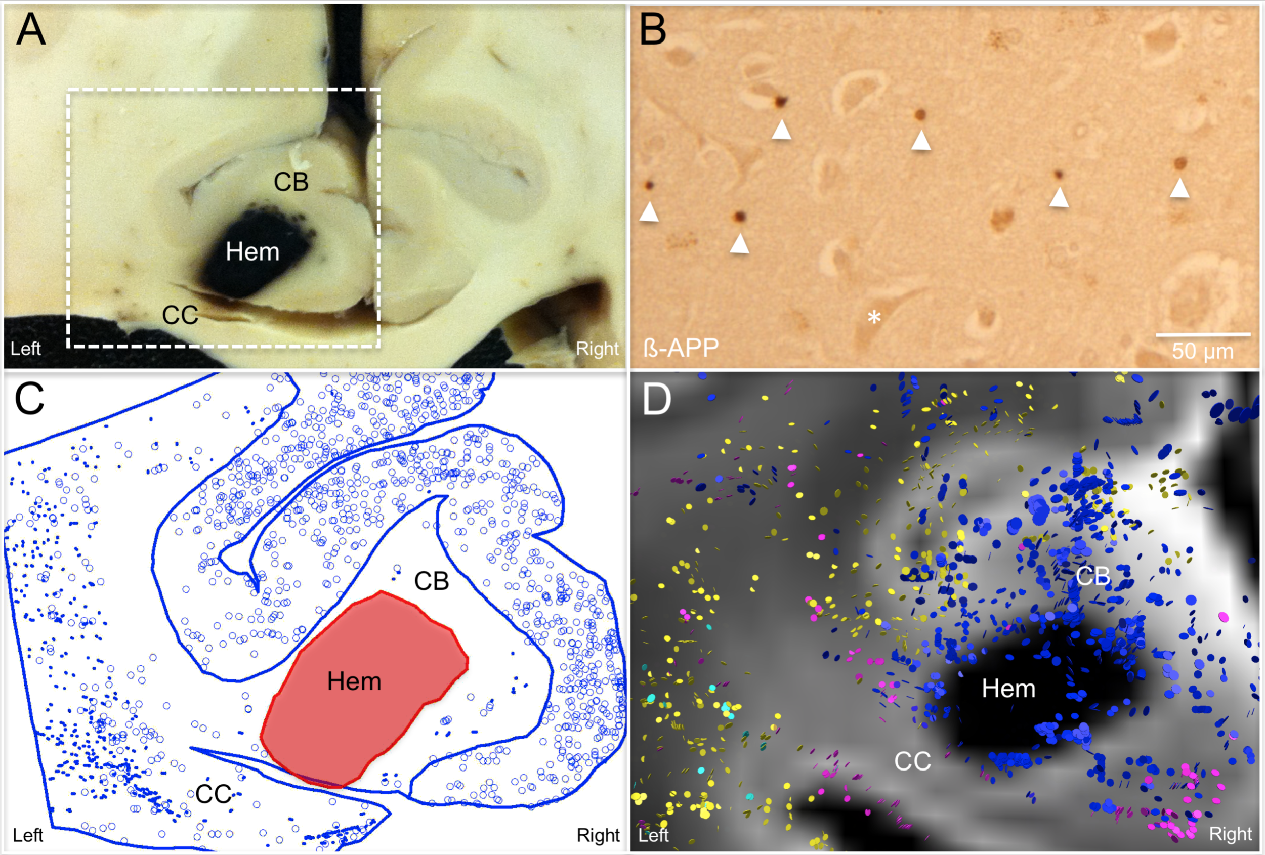
The figure to the right shows a hermorrhage (hem) in the cingulum bundle (CB) and corpus callosum (CC) of a patient who died in the intensive care unit three days after experiencing a severe traumatic brain injury (panel A). Injured axons in the region of the hemorrhage were identified using a beta-amyloid precursor protein immunostain (B-APP), as indicated by the arrow heads in panel B. The full burden of axonal injury is shown in the Neurolucida analysis in panel C, where each dot represents an injured axon. We also performed tractography using postmortem diffusion MRI data to identify injured axonal bundles (panel D). The tractography data show a similar distribution of axonal injury, with additional information provided about the neuroanatomic identity of each injured axonal bundle: blue = injured CB axons; pink = injured CC axons; turquoise = injured thalamocortical axons; yellow = injured axons of undetermined anatomic identity.
This brainstem was scanned using an ultra-high resolution MRI technique before it was sectioned and stained for histopathological analysis. The MRI scan showed severe disruption of brainstem pathways in the coma patient (panel C), as compared to the intact pathways seen in a human control subject (panel D). Microscopic analysis of the patient's brainstem showed severe traumatic axonal injury (panel B, arrowheads), corresponding to sites of fiber tract disruption that were identified by postmortem MRI. Figure adapted from Edlow BL, Haynes RL, Takahashi E, Klein JP, Cummings P, Benner T, Greer DM, Greenberg SM, Wu O, Kinney HC*, Folkerth RD*. Disconnection of the ascending arousal system in traumatic coma. Journal of Neuropathology and Experimental Neurology. 2013;72:505-523. (*co-senior authors).
Selected Ex Vivo Connectomics Publications
Edlow BL, Takahashi E, Wu O, Benner T, Dai G, Bu L, Grant PE, Greer DM, Greenberg SM, Kinney HC, Folkerth RD. Neuroanatomic connectivity of the human ascending arousal system critical to consciousness and its disorders. Journal of Neuropathology and Experimental Neurology. 2012;71:531-546. PMCID PMC3387430. [PubMed] [Altmetric]
Edlow BL, Haynes RL, Takahashi E, Klein JP, Cummings P, Benner T, Greer DM, Greenberg SM, Wu O, Kinney HC*, Folkerth RD*. Disconnection of the ascending arousal system in traumatic coma. Journal of Neuropathology and Experimental Neurology. 2013;72:505-523. (*co-senior authors) PMCID PMC3761353. [PubMed] [Altmetric]
McNab JA*, Edlow BL*, Witzel T, Huang SY, Bhat H, Heberlein K, Feiweier T, Liu K, Keil B, Cohen-Adad J, Tisdall MD, Folkerth RD, Kinney HC, Wald LL. The human connectome project and beyond: Initial applications of 300 mT/m gradients. NeuroImage. 2013;80:234-245. (*co-first authors) PMCID PMC3812060. [PubMed]